Wound Measurement |
Case StudiesDocuments and Links
NPL Calibration Report MAVIS SoftwareDownload (28 MB)Installation Intructions |
|
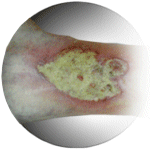
The problem of chronic woundsWounds are familiar to all of us; they are places on the body where the skin has been broken and for most healthy people, healing is rapid and the wound is soon forgotten. However in some circumstances, wounds may become so deep that they resemble craters such as this typical leg wound. These wounds may heal very slowly so that ineffective treatments may be prolonged when a new approach is needed. Clinicians need an objective method of deciding if a wound is getting smaller and therefore healing. The easiest approach is to measure the wound area and volume weekly. |
||
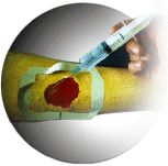
Current wound measurement techniquesThe current method of measuring area is to place a transparent acetate sheet onto the wound and to trace its perimeter. The tracing is then placed onto graph paper and the number of squares counted. It will come as no surprise that this method is inaccurate and unreliable. There are two main methods of measuring the volume of a wound. The first is to fill the wound with saline. The volume dispensed from the syringe equals the wound volume. The main source of error is that the wound absorbs the saline and it is difficult to ensure that the plastic cover takes up the shape of the original healthy skin. The second method is to fill the wound with an alginate or silicone based paste and weigh the amount of material used. |
||
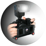 |
THE MAVIS-II Wound Measurement DeviceMAVIS-II is based on the principle of stereo-photogrammetry: note the dual lens system for taking stereo-photographs and the two tube-shaped projectors at the base. These projectors produce two beams of light. When the camera is held at the right angle and distance to the wound these beams of light intersect to form a single spot of light and an image pair can be taken. MAVIS-II is able to:
|
|
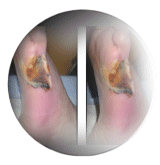 |
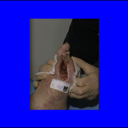
Example of a Wound MeasurementThe MAVIS 3D camera captures 2 images of the wound at the same time. Both images of this excision site on a diabetic foot are virtually identical but show a slight displacement both horizontally and vertically. The vertical displacement is irrelevant but the horizontal displacement of image elements encodes the depth information. (Image courtesy of Dr. Philip Chan, Sheffield Northern General Hospital)
|
|
 |
ResultsAfter manually tracing the perimeter of the wound on screen the wound volume
is found by integrating the distance between the base of the wound and
a minimal surface attached to the wound edges (imagine a volume of
water trapped between the wound bed and a piece of cling film attached
tightly to the wound edge). All results are stored in a data base and can be recalled in various formats. |
|